© 2020 Optical Society of America
Abstract
Acousto-optic imaging (AOI) is a non-invasive method that uses acoustic modulation to map the light fluence inside biological tissue. In many AOI implementations, ultrasound pulses are used in a time-gated measurement to perform depth-resolved imaging without the need for mechanical scanning. However, to achieve high axial resolution, it is required that ultrasound pulses with few cycles are used, limiting the modulation strength. In this Letter, we develop a new approach to pulse-based AOI in which coded ultrasound transmission is used. In coded-transmission AOI (CT-AOI), one may achieve an axial resolution that corresponds to a single cycle, but with a signal-to-noise ratio (SNR) that scales as the square root of the number of cycles. Using CT-AOI with 79 cycles, we experimentally demonstrate over four-fold increase in SNR in comparison to a single-cycle AOI scheme.
One of the fundamental limitations of optical imaging of biological tissue is light scattering due to optical heterogeneity. At depths exceeding several transport lengths, scattering leads to the diffusion of light, which severely limits the imaging resolution that may be achieved [1]. Additionally, optical imaging with diffused light often requires solving nonlinear optimization problems in order to map tissue parameters.
Acousto-optic imaging (AOI) is a hybrid approach that overcomes the limitations of light diffusion by using acoustic modulation [2]. Conventionally, AOI is performed by illuminating the tissue with a highly coherent continuous-wave (CW) laser and using ultrasound to locally modulate the phase of the laser light inside the tissue. In AOI, the ultrasound-induced phase modulation is a result of two mechanisms [3]: pressure-induced modulation of the refractive index and periodic movement of the optical scatterers. When the coherence length of the laser is sufficiently long, the local ultrasound-induced phase modulation inside the tissue is translated into an intensity modulation of the speckle pattern on the tissue boundary. Thus, by measuring the modulation depth of the speckle on the tissue boundary, it is possible to quantify the light fluence within the tissue at the positions in which the acoustic modulation was performed [4].
AOI is capable of identifying both highly absorbing and highly scattering structures through their effect on the light fluence [5], facilitating applications such as early assessment of osteoporosis [6]. Additionally, AOI can provide information on blood flow in the acoustically modulated regions through analysis of the spectral broadening of the speckle modulation [7]. While in most applications AOI is used as an independent technique for assessing tissue parameters, it may also be used as a complimentary technique to optoacoustic tomography (OAT). In previous works [8,9], it has been shown that the information provided by AOI can remove the bias in OAT images due to light attenuation, thus enabling OAT-image quantification.

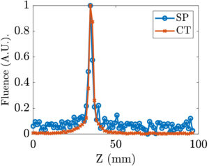
Fig. 1D profile of the modulated light along the ultrasound propagation path for single-pulse AOI (blue) and CT-AOI (red) with corresponding FWMH values of 4.32 mm and 4.07 mm, respectively.
Ahiad Levi, Sagi Monin, Evgeny Hahamovich, Aner Lev, Bruno G. Sfez, and Amir Rosenthal
© 2020 Optical Society of America – Vol. 45, Issue 10, pp. 2858-2861 (2020)


