Biomedical Optics Express Vol. 13, Issue 12, pp. 6229-6244 (2022)
© 2022 Optica Publishing Group under the terms of the Optica Open Access Publishing Agreement
Michael Nagli, Jürgen Koch, Yoav Hazan, Oleg Volodarsky, Resmi Ravi Kumar, Ahiad Levi, Evgeny Hahamovich, Orna Ternyak, Ludger Overmeyer, and Amir Rosenthal
Abstract
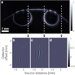
One of the main challenges in miniaturizing optoacoustic technology is the low sensitivity of sub-millimeter piezoelectric ultrasound transducers, which is often insufficient for detecting weak optoacoustic signals. Optical detectors of ultrasound can achieve significantly higher sensitivities than their piezoelectric counterparts for a given sensing area but generally lack acoustic focusing, which is essential in many minimally invasive imaging configurations. In this work, we develop a focused sub-millimeter ultrasound detector composed of a silicon-photonics optical resonator and a micro-machined acoustic lens. The acoustic lens provides acoustic focusing, which, in addition to increasing the lateral resolution, also enhances the signal. The developed detector has a wide bandwidth of 84 MHz, a focal width smaller than 50 µm, and noise-equivalent pressure of 37 mPa/Hz1/2 – an order of magnitude improvement over conventional intravascular ultrasound. We show the feasibility of the approach and the detector’s imaging capabilities by performing high-resolution optoacoustic microscopy of optical phantoms with complex geometries.
[Read more…]
Image generated by GPL Ghostscript (device=ppmraw)
Fig. The detector’s fabrication process. (a) Schematic description of the bonding and the substrate etching steps. (b) A waveguide array fabricated on top of an SOI die (left), an acoustic lens within a quartz substrate (center), and an etched waveguide array bonded to the acoustic lens (right). (c) Fiber bonding setup. The detector is placed under a microscope and between two rotating fiber holders, each connected to a 5-degree of freedom manipulator (x, y, z, pitch, yaw). (d) The assembled detector mounted on the scanning system (3D stage) inside a water tank.
Michael Nagli, Jürgen Koch, Yoav Hazan, Oleg Volodarsky, Resmi Ravi Kumar, Ahiad Levi, Evgeny Hahamovich, Orna Ternyak, Ludger Overmeyer, and Amir Rosenthal.
Biomedical Optics Express Vol. 13, Issue 12, pp. 6229-6244 (2022) •https://doi.org/10.1364/BOE.470295
© 2022 Optica Publishing Group under the terms of the Optica Open Access Publishing Agreement