Michael Nagli, Ron Moisseev, Nathan Suleymanov, Eitan Kaminski, Yoav Hazan, Gil Gelbert, Ilya Goykhman, Amir Rosenthal
Abstract:
Silicon photonics is an emerging platform for acoustic sensing, offering exceptional miniaturization and sensitivity. While efforts have focused on silicon-based
resonators,
silicon nitride resonators can potentially achieve higher Q-factors, further enhancing sensitivity. In this work, a 30 µm
silicon nitride microring resonator was fabricated and coated with an
elastomer to optimize acoustic sensitivity and signal fidelity. The resonator was characterized acoustically, and its capability for optoacoustic
tomography was demonstrated. An acoustic bandwidth of 120 MHz and a noise-equivalent pressure of ∼ 7 mPa/Hz
1/2 were demonstrated. The spatially dependent impulse response agreed with theoretical predictions, and spurious acoustic signals, such as
reverberations and surface
acoustic waves, had a marginal impact. High image fidelity optoacoustic
tomography of a 20 µm knot was achieved, confirming the detector’s imaging capabilities. The results show that silicon
nitride offers low
signal distortion and high-resolution optoacoustic imaging, proving its versatility for acoustic imaging applications.

Fig.
3D optoacoustic reconstruction of a surgical suture. (a) 2D maximum amplitude projection image of the suture. (b) Photograph of the suture showing the similarity between the reconstruction and the imaged object. (a) and (b) are the same scale. (c) 2D slice along the white dashed line shown in (a); shows the cross-section of the suture and demonstrates the lateral and
axial resolutions.
Read more –Photoacoustics Volume 32, August 2023, 100527
Michael Nagli, Ron Moisseev, Nathan Suleymanov, Eitan Kaminski, Yoav Hazan, Gil Gelbert, Ilya Goykhman, Amir Rosenthal


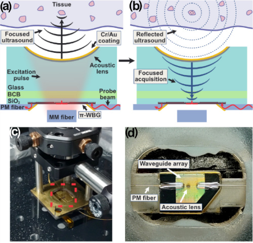
 Fig. Imaging experiments. (a),(c) Optical images of acoustic targets. (b),(d) Ultrasound images of the targets shown in panels (a),(c). (a),(b) Images of 50-µm tungsten wires. (c),(d) Images of lamb meat and fat tissue; (d) is a 1D line scan image along the dotted line shown in panel (c); on the right side are three 0.5 mm × 0.5 mm close-up images. The image in panel (d) is “rolled” and shown in the polar coordinate system.

 Fig. In-vivo Tomographic imaging. Figure 8-a: Microscope images of mouse ear (left) and corresponding MIP of the optoacoustic image (right). Figure 8-b: Montage of four different tomographic depth. The depth difference between each consecutive slice was 50 μm. Figure 8-c. Typical raw OA signals from a mouse ear. Scale bar: 1 mm

 Figure – Normalized power spectrum measured in the spatial position in which the AOI was maximal using conventional ToF-AOI with a PMT (blue) and our homodyne approach with a PD (red). A consistent decrease in noise level measured in both cases in favour of PD. The 2nd and 3rd harmonics of the US signal are visible for both techniques. (Color figure online).