Grüneisen-relaxation photoacoustic microscopy at 1.7 µm and its application in lipid imaging.

Schematic of the GR-PAM experimental system

© 2020 Optical Society of America

Abstract
We report the first, to the best of our knowledge, demonstration of Grüneisen relaxation photoacoustic microscopy (GR-PAM) of lipid-rich tissue imaging at the 1.7 µm band, implemented with a high-energy thulium-doped fiber laser and a fiber-based delay line. GR-PAM enhances the image contrast by intensifying the region of strong absorbers and suppressing out-of-focus signals. Using GR-PAM to image swine-adipose tissue at 1725 nm, an 8.26-fold contrast enhancement is achieved in comparison to conventional PAM. GR-PAM at the 1.7 µm band is expected to be a useful tool for label-free high-resolution imaging of lipid-rich tissue, such as atherosclerotic plaque and nerves.

[Read more…]

Swine muscular tissue images

Fig. Swine muscular tissue images under (a)-(c) OR-PAM and (d)-(f) GR-PAM at (a), (d) 1700; (b), (e) 1725; and (c), (f) 1750 nm. (g) Reference confocal microscopy image at the same region.

Jiawei Shi, Can Li, Huade Mao, Yuxuan Ren, Zhi-Chao Luo, Amir Rosenthal, and Kenneth K. Y. Wong

© 2020 Optical Society of America   Vol. 45, Issue 12, pp. 3268-3271 (2020)

Increased SNR in acousto-optic imaging via coded ultrasound transmission.

© 2020 Optical Society of America

Abstract

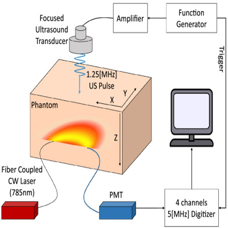
Acousto-optic imaging (AOI) is a non-invasive method that uses acoustic modulation to map the light fluence inside biological tissue. In many AOI implementations, ultrasound pulses are used in a time-gated measurement to perform depth-resolved imaging without the need for mechanical scanning. However, to achieve high axial resolution, it is required that ultrasound pulses with few cycles are used, limiting the modulation strength. In this Letter, we develop a new approach to pulse-based AOI in which coded ultrasound transmission is used. In coded-transmission AOI (CT-AOI), one may achieve an axial resolution that corresponds to a single cycle, but with a signal-to-noise ratio (SNR) that scales as the square root of the number of cycles. Using CT-AOI with 79 cycles, we experimentally demonstrate over four-fold increase in SNR in comparison to a single-cycle AOI scheme.

One of the fundamental limitations of optical imaging of biological tissue is light scattering due to optical heterogeneity. At depths exceeding several transport lengths, scattering leads to the diffusion of light, which severely limits the imaging resolution that may be achieved [1]. Additionally, optical imaging with diffused light often requires solving nonlinear optimization problems in order to map tissue parameters.

Acousto-optic imaging (AOI) is a hybrid approach that overcomes the limitations of light diffusion by using acoustic modulation [2]. Conventionally, AOI is performed by illuminating the tissue with a highly coherent continuous-wave (CW) laser and using ultrasound to locally modulate the phase of the laser light inside the tissue. In AOI, the ultrasound-induced phase modulation is a result of two mechanisms [3]: pressure-induced modulation of the refractive index and periodic movement of the optical scatterers. When the coherence length of the laser is sufficiently long, the local ultrasound-induced phase modulation inside the tissue is translated into an intensity modulation of the speckle pattern on the tissue boundary. Thus, by measuring the modulation depth of the speckle on the tissue boundary, it is possible to quantify the light fluence within the tissue at the positions in which the acoustic modulation was performed [4].

AOI is capable of identifying both highly absorbing and highly scattering structures through their effect on the light fluence [5], facilitating applications such as early assessment of osteoporosis [6]. Additionally, AOI can provide information on blood flow in the acoustically modulated regions through analysis of the spectral broadening of the speckle modulation [7]. While in most applications AOI is used as an independent technique for assessing tissue parameters, it may also be used as a complimentary technique to optoacoustic tomography (OAT). In previous works [8,9], it has been shown that the information provided by AOI can remove the bias in OAT images due to light attenuation, thus enabling OAT-image quantification.

[Read more…]

1D profile of the modulated light along the ultrasound propagation path for single-pulse.

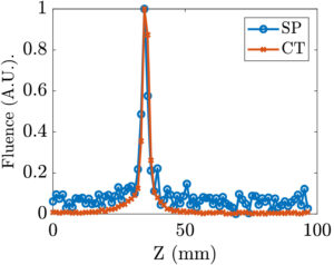
Fig. 1D profile of the modulated light along the ultrasound propagation path for single-pulse AOI (blue) and CT-AOI (red) with corresponding FWMH values of 4.32 mm and 4.07 mm, respectively.


Ahiad Levi, Sagi Monin, Evgeny Hahamovich, Aner Lev, Bruno G. Sfez, and Amir Rosenthal

© 2020 Optical Society of America Vol. 45, Issue 10, pp. 2858-2861 (2020)

Ultrasound Detection Arrays Via Coded Hadamard Apertures.

Experimental setup.

© 2020 IEEE

Abstract
In the medical fields, ultrasound detection is often performed with piezoelectric arrays that enable one to simultaneously map the acoustic fields at several positions. In this work, we develop a novel method for transforming a single-element ultrasound detector into an effective detectionarray by spatially filtering the incoming acoustic fields using a binary acoustic mask coded with cyclic Hadamard patterns. By scanning the mask in front of the detector, we obtain a multiplexed measurement dataset from which a map of the acoustic field is analyticallyconstructed. We experimentally demonstrate our method by transforming a single-element ultrasound detector into 1D arrays with up to 59 elements.

[Read more…]

Fig. Normalized waveform shape comprasion of the strongest signal for a single vs. multiplexed aperture detection. The plots are for (a) an aperture with 1.5 mm diameter and 31 elements and (b) an aperture with 1 mm diameter and 59 elements. 

E. Hahamovich and A. Rosenthal, “Ultrasound Detection Arrays Via Coded Hadamard Apertures,” in IEEE Transactions on Ultrasonics, Ferroelectrics, and Frequency Control, doi: 10.1109/TUFFC.2020.2993583.

© 2020 IEEE

Simultaneous multi-channel ultrasound detection via phase modulated pulse interferometry.

The acoustic setup used to test the performance of PM-PI.

© 2019 Optics Express, OSA publishing.

Abstract
In optical detection of ultrasound, resonators with high Q-factors are often used to maximize sensitivity. However, in order to perform parallel interrogation, conventional interferometric techniques require an overlap between the spectra of all the resonators, which is difficult to achieve with high Q-factor resonators. In this paper, a new method is developed for parallel interrogation of optical resonators with non-overlapping spectra. The method is based on a phase-modulation scheme for pulse interferometry (PM-PI) and requires only a single photodetector and sampling channel per ultrasound detector. Using PM-PI, parallel ultrasound detection is demonstrated with four high Q-factor resonators.

[Read more…]

A schematic drawing of the PM-PI.

Fig. A schematic drawing of the PM-PI system used in this work to interrogate 4 resonators, implemented with π-phase shifted fiber Bragg gratings (π-FBGs). A wideband pulse laser with band-pass filters (BPFs) and an erbium-doped fiber amplifier (EDFA) create a source with a high spectral power density and sufficient bandwidth to cover the spectra of all the resonators. The modulation unit is an unbalanced Mach-Zehnder interferometer (MZI), composed of optical fiber couplers (FC) and a phase modulator (PM). The input phase signal to the PM, shown in the top-right plot, alternates between two values with a difference of 𝜋/2. For each phase value, the pulses interfere differently at the output of each resonator depending on the phase difference in the MZI for the specific resonance wavelength of that resonator. The bottom-right plot, shows a typical voltage signal measured for one of the resonators, which alternates between two states that correspond to the two phase values. As the bottom-right plot shows, in the current implementation, the duration of each phase value delivered to the PM corresponded to 5 laser pulses. We note that the limited bandwidth of our measurement did not allow full separation between the pulses in the bottom-right plot.

Yoav Hazan and Amir Rosenthal. 
Optics Express Vol. 27, Issue 20, pp. 28844-28854 (2019) https://doi.org/10.1364/OE.27.028844


Enhanced Sensitivity of Silicon-Photonics-Based Ultrasound Detection via BCB Coating.

Sio and Bcb over clading

© 2019 IEEE Photonics Journal

Impact Statement:
This paper gives a solution to one of the fundamental limitations of silicon-photonics based ultrasound detectors: the low photo-elastic response of silicon and silica. By using a BCB over-cladding, 5-fold increase in the acoustic sensitivity is achieved. We additionally provide a detailed analysis of the sensing mechanism, quantifying the different effects that contribute to the enhanced sensitivity.
Abstract:
Ultrasound detection via silicon waveguides relies on the ability of acoustic waves to modulate the effective refractive index of the guided modes. However, the low photo-elastic response of silicon and silica limits the sensitivity of conventional silicon-on-insulator sensors, in which the silicon core is surrounded by a silica cladding. In this paper, we demonstrate that the sensitivity of silicon waveguides to ultrasound may be significantly enhanced by replacing the silica over-cladding with bisbenzocyclobutene (BCB)-a transparent polymer with a high photo-elastic coefficient. In our experimental study, the response to ultrasound, in terms of the induced modulation in the effective refractive index, achieved for a BCB-coated silicon waveguide with TM polarization was comparable to values previously reported for polymer waveguides and an order of magnitude higher than the response achieved by an optical fiber. In addition, in our study, the susceptibility of the sensors to surface acoustic waves and reverberations was reduced for both TE and TM modes when the BCB over-cladding was used.

[Read more…]

setup schematic

Fig. – The measurement setup. For each of the polarizations, a Mach-Zehnder interferometer was constructed, where in each of the interferometer arms a chip with a different over-cladding material (BCB or silica) was connected. An ultrasound transducer was used to generate acoustic waves that impinged on only one of the chips, which were separated by more than 10 cm.

Resmi Ravi Kumar, Evgeny Hahamovich, Shai Tsesses, Yoav Hazan, Assaf Grinberg, Amir Rosenthal. IEEE Photonics Journal ( Volume: 11 , Issue: 3 , June 2019 )

Noise reduction in resonator-based ultrasound sensors by using a CW laser and phase detection.

© 2019 Optical Society of America

The detection of ultrasound via optical resonators is conventionally performed by tuning a continuous-wave (CW) laser to the linear slope of the resonance and monitoring the intensity modulation at the resonator output. While intensity monitoring offers the advantage of simplicity, its sensitivity is often limited by the frequency noise of the CW laser. In this work, we develop an alternative CW technique that can significantly reduce measurement noise by monitoring variations in the phase, rather than intensity, at the resonator output. In our current implementation, which is based on a balanced Mach–Zehnder interferometer for phase detection, we demonstrate a 24-fold increase in the signal-to-noise ratio of the detected ultrasound signal over the conventional, intensity-monitoring approach.[Read more…]

Signals detected using the IM and PM scheme

Fig. – Signals detected using the IM and PM interrogation methods with an optical power of 0.1 mW. In the PM measurement, the OPD was set to zero to maximize the SNR.

Lucas Riobó, Yoav Hazan, Francisco Veiras, María Garea, Patricio Sorichetti, and Amir Rosenthal.  Optics Letters Vol. 44, Issue 11, pp. 2677-2680 (2019) •https://doi.org/10.1364/OL.44.002677
© 2019 Optical Society of America

The Impulse Response of Negatively Focused Spherical Ultrasound Detectors and its Effect on Tomographic Optoacoustic Reconstruction.

The Impulse Response

source: © 2019  IEEE Transactions on Medical Imaging. 

In optoacoustic tomography, negatively focused detectors have been shown to improve the tangential image resolution without sacrificing sensitivity. Since no exact inversion formulae exist for optoacoustic image reconstruction with negatively focused detectors, image reconstruction in such cases is based on using the virtual-detector approximation, in which it is assumed that the response of the negatively focused detector is identical, up to a constant time delay, to that of a point-like detector positioned in the detector’s center of curvature. In this work, we analyze the response of negatively focused spherical ultrasound detectors in three dimensions and demonstrate how their properties affect the optoacoustic reconstruction. Our analysis sheds new light on commonly reported experimental reconstruction artifacts in optoacoustic systems that employ negatively focused detectors. Based on our analysis, we introduce a simple correction to the virtual-detector approximation that significantly enhances image contrast and reduces artifacts.  [Read more…]

The Impulse Response

Fig. (a) The geometry of the negative acoustic lens studied with full
acoustic simulations. The speed of sound of the surrounding medium and lens
material were 1500 m/s and 2757 m/s. respectively. (b) The detected acoustic
signals obtained when the lens was acoustically matched to the surrounding
medium (solid-blue curve) and when its acoustic impedance was 1.xx times that
of the surrounding medium (dashed-red curve), leading to internal reflections
in the lens structure. The reflection from the detection surface was 50% of the
pressure signal. 

Gilad Drozdov, Ahiad Levi, Amir Rosenthal.
IEEE Transactions on Medical Imaging.DOI: 10.1109/TMI.2019.2897588


Everolimus-eluting stents stabilize plaque inflammation in vivo: assessment by intravascular fluorescence molecular imaging.

source: © 2016 European Heart Journal

Aims
Inflammation drives atherosclerosis complications and is a promising therapeutic target for plaque stabilization. At present, it is unknown whether local stenting approaches can stabilize plaque inflammation in vivo. Here, we investigate whether everolimus-eluting stents (EES) can locally suppress plaque inflammatory protease activity in vivo using intravascular near-infrared fluorescence (NIRF) molecular imaging.
Methods and results
Balloon-injured, hyperlipidaemic rabbits with atherosclerosis received non-overlapping EES and bare metal stents (BMS) placement into the infrarenal aorta (n = 7 EES, n = 7 BMS, 3.5 mm diameter x 12 mm length). Four weeks later, rabbits received an injection of the cysteine protease-activatable NIRF imaging agent Prosense VM110. Twenty-four hours later, co-registered intravascular 2D NIRF, X-ray angiography and intravascular ultrasound imaging were performed. In vivo EES-stented plaques contained substantially reduced NIRF inflammatory protease activity compared with untreated plaques and BMS-stented plaques (P = 0.006). Ex vivo macroscopic NIRF imaging of plaque protease activity corroborated the in vivo results (P = 0.003). Histopathology analyses revealed that EES-treated plaques showed reduced neointimal and medial arterial macrophage and cathepsin B expression compared with unstented and BMS-treated plaques.
Conclusions
EES-stenting stabilizes plaque inflammation as assessed by translational intravascular NIRF molecular imaging in vivo. These data further support that EES may provide a local approach for stabilizing inflamed plaques.[Read more…]

FRI of plaque

Fig. Ex vivo FRI analyses of plaque inflammatory cysteine protease activity in BMS-, EES-treated, and unstented plaque zones. (A–C) Ex vivo FRI alignments from three representative animals. All NIRF images were obtained with a one second exposure. Image windows optimized for individual images.

Marcella A. Calfon Press, Georgios Mallas, Amir Rosenthal, Tetsuya Hara, Adam Mauskapf, R. Nika Nudelman, Alexander Sheehy, Igor V. Polyakov, Frank Kolodgie, Renu Virmani, J. Luis Guerrero, Vasilis Ntziachristos, Farouc A. Jaffer.European Heart Journal – Cardiovascular Imaging, Volume 18, Issue 5, 1 May 2017, Pages 510–518.

Optoacoustic image reconstruction and system analysis for finite-aperture detectors under the wavelet-packet framework.

Schematic illustration

source: © 2016 J. of Biomedical Optics

In optoacoustic tomography, detectors with relatively large areas are often employed to achieve high detection sensitivity. However, spatial-averaging effects over large detector areas may lead to attenuation of high acoustic frequencies and, subsequently, loss of fine features in the reconstructed image. Model-based reconstruction algorithms improve image resolution in such cases by correcting for the effect of the detector’s aperture on the detected signals. However, the incorporation of the detector’s geometry in the optoacoustic model leads to a significant increase of the model matrix memory cost, which hinders the application of inversion and analysis tools such as singular value decomposition (SVD). We demonstrate the use of the wavelet-packet framework for optoacoustic systems with finite-aperture detectors. The decomposition of the model matrix in the wavelet-packet domain leads to sufficiently smaller model matrices on which SVD may be applied. Using this methodology over an order of magnitude reduction in inversion time is demonstrated for numerically generated and experimental data. Additionally, our framework is demonstrated for the analysis of inversion stability and reveals a new, nonmonotonic dependency of the system condition number on the detector size. Thus, the proposed framework may assist in choosing the optimal detector size in future optoacoustic systems.[Read more…]

Optoacoustic reconstructions of a mouse

Fig.Optoacoustic reconstructions of a mouse’s head from limited view (180 deg) experimental data obtained using (a) BP, (b) IMMI, (c) IMMI-FAD, and (d) GWP-IMMI-FAD.

Yiyong Han, Vasilis Ntziachristos, Amir Rosenthal. J. of Biomedical Optics, 21(1), 016002 (2016). https://doi.org/10.1117/1.JBO.21.1.016002

High-Throughput Sparsity-Based Inversion Scheme for Optoacoustic Tomography

source: © 2016 IEEE Transactions on Medical Imaging

The concept of sparsity is extensively exploited in the fields of data acquisition and image processing, contributing to better signal-to-noise and spatio-temporal performance of the various imaging methods. In the field of optoacoustic tomography, the image reconstruction problem is often characterized by computationally extensive inversion of very large datasets, for instance when acquiring volumetric multispectral data with high temporal resolution. In this article we seek to accelerate accurate model-based optoacoustic inversions by identifying various sources of sparsity in the forward and inverse models as well as in the single- and multi-frame representation of the projection data. These sources of sparsity are revealed through appropriate transformations in the signal, model and image domains and are subsequently exploited for expediting image reconstruction. The sparsity-based inversion scheme was tested with experimental data, offering reconstruction speed enhancement by a factor of 40 to 700 times as compared with the conventional iterative model-based inversions while preserving similar image quality. The demonstrated results pave the way for achieving real-time performance of model-based reconstruction in multi-dimensional optoacoustic imaging.[Read more…..]

Fingers us samples

Fig.3d maximum intensity projections of the volumetric dataset showing a 3d angiogram of a human finger obtained by cross-sectional scan in the z direction. (a) Photograph of the finger and reconstructions obtained with (b) LSQR-15, (c) WP-o, and (d) PCA-wp-T are shown.

Christian Lutzweiler, Stratis Tzoumas, Amir Rosenthal, Vasilis Ntziachristos, Daniel Razansky. Published in: IEEE Transactions on Medical Imaging ( Volume: 35 , Issue: 2 , Feb. 2016 ).