


 Fig. PA image of (a) water acquired by 1930-nm OR-PAM and b lipid acquired by 1750-nm OR-PAM; c Overlaid PA image of (a) and (b). d Photography of the adipose tissue. e–f Three-dimensional rendering view of (a, b). Scale bars, 500 µm
Read more – eLight 2, 6 (2022)
Jiawei Shi, Mingsheng Li, Huajun Tang, Jiqiang Kang, Najia Sharmin, Amir Rosenthal & Kenneth K. Y. Wong
Fig. PA image of (a) water acquired by 1930-nm OR-PAM and b lipid acquired by 1750-nm OR-PAM; c Overlaid PA image of (a) and (b). d Photography of the adipose tissue. e–f Three-dimensional rendering view of (a, b). Scale bars, 500 µm
Read more – eLight 2, 6 (2022)
Jiawei Shi, Mingsheng Li, Huajun Tang, Jiqiang Kang, Najia Sharmin, Amir Rosenthal & Kenneth K. Y. Wong
Hybrid optical parametrically-oscillating emitter at 1930 nm for volumetric photoacoustic imaging of water content

eLight 2, 6 (2022)
Jiawei Shi, Mingsheng Li, Huajun Tang, Jiqiang Kang, Najia Sharmin, Amir Rosenthal & Kenneth K. Y. Wong
Abstract:
Water plays a vital role in biological metabolism and it would be essential to trace the water content non-invasively, such as leveraging the vibrational absorption peak of the O–H bond. However, due to the lack of an efficient laser source, it was challenging to image the water content in the deep tissue with micron-level spatial resolution. To address this problem, we develop a high-power hybrid optical parametrically-oscillating emitter (HOPE) at 1930 nm, at which the vibrational absorption peak of the O–H bond locates. The maximum pulse energy is over 1.74 μJ with a pulse repetition rate of 50 kHz and a pulse width of 15 ns. We employ this laser source in the optical-resolution photoacoustic microscopy (OR-PAM) system to image the water content in the phantom and the biological tissue in vitro. Our 1930-nm OR-PAM could map the water content in the complex tissue environment at high spatial resolution, deep penetration depth, improved sensitivity, and suppressed artifact signal of the lipid.
Fig. PA image of (a) water acquired by 1930-nm OR-PAM and b lipid acquired by 1750-nm OR-PAM; c Overlaid PA image of (a) and (b). d Photography of the adipose tissue. e–f Three-dimensional rendering view of (a, b). Scale bars, 500 µm
Read more – eLight 2, 6 (2022)
Jiawei Shi, Mingsheng Li, Huajun Tang, Jiqiang Kang, Najia Sharmin, Amir Rosenthal & Kenneth K. Y. Wong


 The study was supported by the Russell Berrie Nanotechnology Institute (RBNI), the National Science Foundation, the Polak Foundation, the Israel Innovation Authority, the Israel Science Foundation and the Ollendorf Minerva Center.
 Figure-Optoacoustic images of the blood vessels in a human wrist, at different depths and orientations. A cross-section of the radial artery can be seen clearly in real time at depths up to 7 mm, as in (a,c). A deep vein can be seen in (b) at a depth of 8 mm. In (d), we can see a vein diving from 3 to 7 mm in a longitudinal cross-section. The scale bar in subfigure (a) applies to all subfigures.
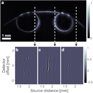


 Fig. PTAM pressure measurements. (a) Transmission spectrum of π-BG at different static pressures. The legend notes the pressure in kPa. (b) Normalized power transmission, (c) peak transmission, (d) resonance width, and (e) resonance wavelength of π-BG at different pressure calculated from the spectrum plotted in panel (a). (f) Schematic configuration of simultaneous ultrasound signal detection. Ultrasound signal, generated by a transducer, impinges the detection array at an angle. The setup results in a slight delay difference of the ultrasound signal along the detector array. (g) and (h) Measured ultrasound signals of the setup in panel (f), for resonators presented in Figs. 1(e) and 1(f), respectively.

 Fig. A is a photograph of the leg. B is a subset over a vertical line of the measured signals, C is the de-multiplexed signals, and D is the MAP of the reconstructed optical density as a function of depth (z). The amplitudes are in arbitrary units.

