2023 Optics Letters Vol. 48, Issue 10, pp. 2668-2671
Michael Nagli, Jürgen Koch, Yoav Hazan, Ahiad Levi, Orna Ternyak, Ludger Overmeyer, and Amir Rosenthal
Abstract
We present an all-optical focused ultrasound transducer with a sub-millimeter aperture and demonstrate its capability for high-resolution imaging of tissue ex vivo. The transducer is composed of a wideband silicon photonics ultrasound detector and a miniature acoustic lens coated with a thin optically absorbing metallic layer used to produce laser-generated ultrasound. The demonstrated device achieves axial resolution and lateral resolutions of 12 μm and 60 μm, respectively, well below typical values achieved by conventional piezoelectric intravascular ultrasound. The size and resolution of the developed transducer may enable its use for intravascular imaging of thin fibrous cap atheroma.
[Read more…]
Fig. Imaging experiments. (a),(c) Optical images of acoustic targets. (b),(d) Ultrasound images of the targets shown in panels (a),(c). (a),(b) Images of 50-µm tungsten wires. (c),(d) Images of lamb meat and fat tissue; (d) is a 1D line scan image along the dotted line shown in panel (c); on the right side are three 0.5 mm × 0.5 mm close-up images. The image in panel (d) is “rolled” and shown in the polar coordinate system.
Michael Nagli, Jürgen Koch, Yoav Hazan, Ahiad Levi, Orna Ternyak, Ludger Overmeyer, and Amir Rosenthal
2023 Biomedical Engineering Letters
Tamar Harary, Yoav Hazan & Amir Rosenthal
Abstract
High-resolution optoacoustic imaging at depths beyond the optical diffusion limit is conventionally performed using a microscopy setup where a strongly focused ultrasound transducer samples the image object point-by-point. Although recent advancements in miniaturized ultrasound detectors enables one to achieve microscopic resolution with an unfocused detector in a tomographic configuration, such an approach requires illuminating the entire object, leading to an inefficient use of the optical power, and imposing a trans-illumination configuration that is limited to thin objects. We developed an optoacoustic micro-tomography system in an epi-illumination configuration, in which the illumination is scanned with the detector. The system is demonstrated in phantoms for imaging depths of up to 5 mm and in vivo for imaging the vasculature of a mouse ear. Although image-formation in optoacoustic tomography generally requires static illumination, our numerical simulations and experimental measurements show that this requirement is relaxed in practice due to light diffusion, which homogenizes the fluence in deep tissue layers.
[Read more…]
Fig. In-vivo Tomographic imaging. Figure 8-a: Microscope images of mouse ear (left) and corresponding MIP of the optoacoustic image (right). Figure 8-b: Montage of four different tomographic depth. The depth difference between each consecutive slice was 50 μm. Figure 8-c. Typical raw OA signals from a mouse ear. Scale bar: 1 mm
2023 Biomedical Engineering Letters volume 13, pages49–56
Ahiad R.Levi, Yoav Hazan, Aner Lev, Bruno G. Sfez & Amir Rosenthal
Abstract
Acousto-optics imaging (AOI) is a hybrid imaging modality that is capable of mapping the light fluence rate in deep tissue by local ultrasound modulation of the diffused photons. Since the intensity of the modulated photons is relatively low, AOI systems often rely on high-gain photodetectors, e.g. photomultiplier tubes (PMTs), which limit scalability due to size and cost and may significantly increase the relative shot-noise in the detected signal due to low quantum yields or gain noise. In this work, we have developed a homodyne AOI scheme in which the modulated photons are amplified by interference with a reference beam, enabling their detection with a single low-gain photodetector in reflection-mode configuration. We experimentally demonstrate our approach with a silicon photodiode, achieving over a 4-fold improvement in SNR in comparison to a PMT-based setup. The increased SNR manifested in lower background noise level thus enabling deeper imaging depths. The use of a fiber-based configuration enables the integration of our scheme in a hand-held AOI probe.
[Read more…]
Figure – Normalized power spectrum measured in the spatial position in which the AOI was maximal using conventional ToF-AOI with a PMT (blue) and our homodyne approach with a PD (red). A consistent decrease in noise level measured in both cases in favour of PD. The 2nd and 3rd harmonics of the US signal are visible for both techniques. (Color figure online).
Ahiad R.Levi, Yoav Hazan, Aner Lev, Bruno G. Sfez & Amir Rosenthal
Photonics 2022, 9(12), 907
Zohar Or, Ahiad R.Levi, Yoav Hazan and Amir Rosenthal
Abstract
The ability to rapidly locate blood vessels in patients is important in many clinical applications, e.g., in catheterization procedures. Optical techniques, including visual inspection, generally suffer from a reduced performance at depths below 1 mm, while ultrasound and optoacoustic tomography are better suited to a typical depth on the scale of 1 cm and require an additional spacer between the tissue and transducer in order to image the superficial structures at the focus plane. For this work, we developed a hand-held optoacoustic probe, designed for localizing blood vessels from the contact point down to a depth of 1 cm, without the use of a spacer. The probe employs a flat lens-free ultrasound array, enabling a largely depth-independent response down to a depth of 1 cm, at the expense of low elevational resolution. Specifically, while in lens-based probes, the acoustic signals from outside the focal region suffer from distortion, in our probe, only the amplitude of the signal varies with depth, thus leading to an imaging quality that is largely depth-independent in the imaged region. To facilitate miniaturization, dark-field illumination is used, whereby light scattering from the tissue is exploited to homogenize the sensitivity field.
[Read more…]
Figure-Optoacoustic images of the blood vessels in a human wrist, at different depths and orientations. A cross-section of the radial artery can be seen clearly in real time at depths up to 7 mm, as in (a,c). A deep vein can be seen in (b) at a depth of 8 mm. In (d), we can see a vein diving from 3 to 7 mm in a longitudinal cross-section. The scale bar in subfigure (a) applies to all subfigures.
Zohar Or, Ahiad R.Levi, Yoav Hazan and Amir Rosenthal
Michael Nagli, Jürgen Koch, Yoav Hazan, Oleg Volodarsky, Resmi Ravi Kumar, Ahiad Levi, Evgeny Hahamovich, Orna Ternyak, Ludger Overmeyer, and Amir Rosenthal.
2022 Communications Engineering
Evgeny Hahamovich, Sagi Monin, Ahiad Levi, Yoav Hazan & Amir Rosenthal
Abstract
Optoacoustic tomography (OAT) is a hybrid imaging modality that combines optical excitation with ultrasound detection and enables high-resolution visualization of optical contrasts at tissue depths in which light is completely diffused. Despite its promise in numerous research and clinical applications, OAT is limited by the technological immaturity of ultrasound detection systems. It suffers from limited element count, narrow field of view and lack of technology for spatial modulation of acoustic signals. Here we report single-detector OAT capable of high-fidelity imaging using an amplitude mask in planar geometry coded with cyclic patterns for structured spatial acoustic modulation. Our image reconstruction method maximises sensitivity, is compatible with planar signal detection, and uses only linear operations, thus avoiding artefacts associated with the nonlinear compressed-sensing inversion. We demonstrate our method for 3D OAT of complex objects and living tissue performed with only a single ultrasound detector, effectively coded into a 2D array with 1763 elements. Our method paves the way for a new generation of high-fidelity, low-cost OAT systems.
[Read more…]
Fig. A is a photograph of the leg. B is a subset over a vertical line of the measured signals, C is the de-multiplexed signals, and D is the MAP of the reconstructed optical density as a function of depth (z). The amplitudes are in arbitrary units.
Abstract Optical imaging is commonly performed with either a camera and wide-field illumination or with a single detector and a scanning collimated beam; unfortunately, these options do not exist at all wavelengths. Single-pixel imaging offers an alternative that can be performed with a single detector and wide-field illumination, potentially enabling imaging applications in which the detection and illumination technologies are immature. However, single-pixel imaging currently suffers from low imaging rates owing to its reliance on configurable spatial light modulators, generally limited to 22 kHz rates. We develop an approach for rapid single-pixel imaging which relies on cyclic patterns coded onto a spinning mask and demonstrate it for in vivo imaging of C. elegans worms. Spatial modulation rates of up to 2.4 MHz, imaging rates of up to 72 fps, and image-reconstruction times of down to 1.5 ms are reported, enabling real-time visualization of dynamic objects.
a Video capturing of vertically shifted resolution target, 101 × 103 resolution and 72 fps frame rate. Total recording rate of 0.75 M pixels per second. Frames 1, 30, 60, 90, 110, and 142 out of the 142 captured frames are presented. The red and blue circles mark constant positions on the resolution target. b, c Videos capturing the motion of C. elegans worms at a frame rate of 10 fps, corresponding to a total recording rate of 0.7 M pixels per second. Frames 5, 10, 15, 20, 25, and 30 out of 31 frames are presented.
Abstract Single-pixel imaging (SPI) enables the visualization of objects with a single detector by using a sequence of spatially modulated illumination patterns. For natural images, the number of illumination patterns may be smaller than the number of pixels when compressed-sensing algorithms are used. Nonetheless, the sequential nature of the SPI measurement requires that the object remains static until the signals from all the required patterns have been collected. In this paper, we present a new approach to SPI that enables imaging scenarios in which the imaged object, or parts thereof, moves within the imaging plane during data acquisition. Our algorithms estimate the motion direction from inter-frame cross-correlations and incorporate it in the reconstruction model. Moreover, when the illumination pattern is cyclic, the motion may be estimated directly from the raw data, further increasing the numerical efficiency of the algorithm. A demonstration of our approach is presented for both numerically simulated and measured data.


 The study was supported by the Russell Berrie Nanotechnology Institute (RBNI), the National Science Foundation, the Polak Foundation, the Israel Innovation Authority, the Israel Science Foundation and the Ollendorf Minerva Center.
Read more – © 2022 Nature Comunication -Silicon-photonics acoustic detector for optoacoustic micro-tomography.
The study was supported by the Russell Berrie Nanotechnology Institute (RBNI), the National Science Foundation, the Polak Foundation, the Israel Innovation Authority, the Israel Science Foundation and the Ollendorf Minerva Center.
Read more – © 2022 Nature Comunication -Silicon-photonics acoustic detector for optoacoustic micro-tomography.
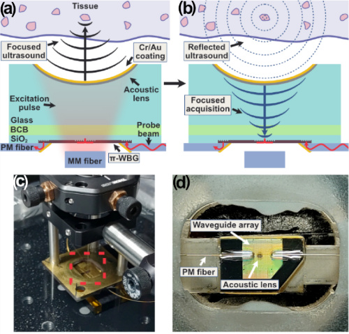
 Fig. Imaging experiments. (a),(c) Optical images of acoustic targets. (b),(d) Ultrasound images of the targets shown in panels (a),(c). (a),(b) Images of 50-µm tungsten wires. (c),(d) Images of lamb meat and fat tissue; (d) is a 1D line scan image along the dotted line shown in panel (c); on the right side are three 0.5 mm × 0.5 mm close-up images. The image in panel (d) is “rolled” and shown in the polar coordinate system.

 Fig. In-vivo Tomographic imaging. Figure 8-a: Microscope images of mouse ear (left) and corresponding MIP of the optoacoustic image (right). Figure 8-b: Montage of four different tomographic depth. The depth difference between each consecutive slice was 50 μm. Figure 8-c. Typical raw OA signals from a mouse ear. Scale bar: 1 mm

 Figure – Normalized power spectrum measured in the spatial position in which the AOI was maximal using conventional ToF-AOI with a PMT (blue) and our homodyne approach with a PD (red). A consistent decrease in noise level measured in both cases in favour of PD. The 2nd and 3rd harmonics of the US signal are visible for both techniques. (Color figure online).
 Figure-Optoacoustic images of the blood vessels in a human wrist, at different depths and orientations. A cross-section of the radial artery can be seen clearly in real time at depths up to 7 mm, as in (a,c). A deep vein can be seen in (b) at a depth of 8 mm. In (d), we can see a vein diving from 3 to 7 mm in a longitudinal cross-section. The scale bar in subfigure (a) applies to all subfigures.
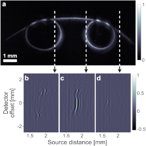


 Fig. PTAM pressure measurements. (a) Transmission spectrum of π-BG at different static pressures. The legend notes the pressure in kPa. (b) Normalized power transmission, (c) peak transmission, (d) resonance width, and (e) resonance wavelength of π-BG at different pressure calculated from the spectrum plotted in panel (a). (f) Schematic configuration of simultaneous ultrasound signal detection. Ultrasound signal, generated by a transducer, impinges the detection array at an angle. The setup results in a slight delay difference of the ultrasound signal along the detector array. (g) and (h) Measured ultrasound signals of the setup in panel (f), for resonators presented in Figs. 1(e) and 1(f), respectively.

 Fig. A is a photograph of the leg. B is a subset over a vertical line of the measured signals, C is the de-multiplexed signals, and D is the MAP of the reconstructed optical density as a function of depth (z). The amplitudes are in arbitrary units.

