Unveiling the evolution of light within photonic integrated circuits.

Fourier-space imaging and extraction of the wave vector components.

Optica Vol. 11, Issue 1, pp. 42-47 (2024)

Matan Iluz, Kobi Cohen, Jacob Kheireddine, Yoav Hazan, Amir Rosenthal, Shai Tsesses, and Guy Bartal

Abstract:

Silicon photonics leverages mature semiconductor technology to produce cost-effective and high-performance components for various applications in data centers, artificial intelligence, and quantum computing. While the geometry of photonic integrated circuits can be characterized by existing means, their optimal and accurate performance requires detailed characterization of the light propagating within them. Here we demonstrate the first, to our knowledge, direct visualization of the light as it travels inside photonic integrated circuits. We employ the natural nonlinear optical properties of silicon to directly map the electric field of the waves guided inside the integrated circuits, characterizing waveguides and multimode splitters while extracting various parameters of the device—all in real-time and in a noninvasive manner. Our approach for visualizing light inside photonic circuits is the only solution directly providing such information without any overhead or penalty, potentially making it a crucial component for the characterization of photonic circuitry, toward their improved design, fabrication, and optimization.

Imaging light within the MMI splitter.

Imaging light within the MMI splitter. (a) Optical imaging of the MMI device. (b) Direct mapping of the light evolution inside an MMI device. The figure comprises exposures of seven different locations along the device, stitched together to track the evolution in the MMI. The rightmost exposure is at the single-waveguide input while the leftmost is of the two waveguides. (c) Zoom-in of (b). (d) Simulation results on a similar scale to (c). (b)–(d) show the intensity of the transverse electric field. Evidently, the experimental results are in excellent agreement with the simulation of light evolution in such a system.
.

Optica Vol. 11, Issue 1, pp. 42-47 (2024)

Matan Iluz, Kobi Cohen, Jacob Kheireddine, Yoav Hazan, Amir Rosenthal, Shai Tsesses, and Guy Bartal

Silicon photonic acoustic detector (SPADE) using a silicon nitride microring resonator.

setup image

Photoacoustics Volume 32, August 2023, 100527

Michael Nagli, Ron Moisseev, Nathan Suleymanov, Eitan Kaminski, Yoav Hazan, Gil Gelbert, Ilya Goykhman, Amir Rosenthal Abstract: Silicon photonics is an emerging platform for acoustic sensing, offering exceptional miniaturization and sensitivity. While efforts have focused on silicon-based resonatorssilicon nitride resonators can potentially achieve higher Q-factors, further enhancing sensitivity. In this work, a 30 µm silicon nitride microring resonator was fabricated and coated with an elastomer to optimize acoustic sensitivity and signal fidelity. The resonator was characterized acoustically, and its capability for optoacoustic tomography was demonstrated. An acoustic bandwidth of 120 MHz and a noise-equivalent pressure of ∼ 7 mPa/Hz1/2 were demonstrated. The spatially dependent impulse response agreed with theoretical predictions, and spurious acoustic signals, such as reverberations and surface acoustic waves, had a marginal impact. High image fidelity optoacoustic tomography of a 20 µm knot was achieved, confirming the detector’s imaging capabilities. The results show that silicon nitride offers low signal distortion and high-resolution optoacoustic imaging, proving its versatility for acoustic imaging applications. silicone_main picture Fig. 3D optoacoustic reconstruction of a surgical suture. (a) 2D maximum amplitude projection image of the suture. (b) Photograph of the suture showing the similarity between the reconstruction and the imaged object. (a) and (b) are the same scale. (c) 2D slice along the white dashed line shown in (a); shows the cross-section of the suture and demonstrates the lateral and axial resolutions. Read more –Photoacoustics Volume 32, August 2023, 100527 Michael Nagli, Ron Moisseev, Nathan Suleymanov, Eitan Kaminski, Yoav Hazan, Gil Gelbert, Ilya Goykhman, Amir Rosenthal

High-resolution silicon photonics focused ultrasound transducer with a sub-millimeter aperture.

Schematic illustration of the developed transducer
2023 Optics Letters Vol. 48, Issue 10, pp. 2668-2671 Michael Nagli, Jürgen Koch, Yoav Hazan, Ahiad Levi, Orna Ternyak, Ludger Overmeyer, and Amir Rosenthal Abstract We present an all-optical focused ultrasound transducer with a sub-millimeter aperture and demonstrate its capability for high-resolution imaging of tissue ex vivo. The transducer is composed of a wideband silicon photonics ultrasound detector and a miniature acoustic lens coated with a thin optically absorbing metallic layer used to produce laser-generated ultrasound. The demonstrated device achieves axial resolution and lateral resolutions of 12 μm and 60 μm, respectively, well below typical values achieved by conventional piezoelectric intravascular ultrasound. The size and resolution of the developed transducer may enable its use for intravascular imaging of thin fibrous cap atheroma. [Read more…] Imaging experiments. Fig. Imaging experiments. (a),(c) Optical images of acoustic targets. (b),(d) Ultrasound images of the targets shown in panels (a),(c). (a),(b) Images of 50-µm tungsten wires. (c),(d) Images of lamb meat and fat tissue; (d) is a 1D line scan image along the dotted line shown in panel (c); on the right side are three 0.5 mm × 0.5 mm close-up images. The image in panel (d) is “rolled” and shown in the polar coordinate system.

Michael Nagli, Jürgen Koch, Yoav Hazan, Ahiad Levi, Orna Ternyak, Ludger Overmeyer, and Amir Rosenthal

2023 Optics Letters Vol. 48, Issue 10, pp. 2668-2671 https://doi.org/10.1364/OL.486567

Silicon-photonics focused ultrasound detector for minimally invasive optoacoustic imaging.

ptoacoustic image of a double-loop-shaped
Biomedical Optics Express Vol. 13, Issue 12, pp. 6229-6244 (2022) © 2022 Optica Publishing Group under the terms of the Optica Open Access Publishing Agreement Michael Nagli, Jürgen Koch, Yoav Hazan, Oleg Volodarsky, Resmi Ravi Kumar, Ahiad Levi, Evgeny Hahamovich, Orna Ternyak, Ludger Overmeyer, and Amir Rosenthal Abstract One of the main challenges in miniaturizing optoacoustic technology is the low sensitivity of sub-millimeter piezoelectric ultrasound transducers, which is often insufficient for detecting weak optoacoustic signals. Optical detectors of ultrasound can achieve significantly higher sensitivities than their piezoelectric counterparts for a given sensing area but generally lack acoustic focusing, which is essential in many minimally invasive imaging configurations. In this work, we develop a focused sub-millimeter ultrasound detector composed of a silicon-photonics optical resonator and a micro-machined acoustic lens. The acoustic lens provides acoustic focusing, which, in addition to increasing the lateral resolution, also enhances the signal. The developed detector has a wide bandwidth of 84 MHz, a focal width smaller than 50 µm, and noise-equivalent pressure of 37 mPa/Hz1/2 – an order of magnitude improvement over conventional intravascular ultrasound. We show the feasibility of the approach and the detector’s imaging capabilities by performing high-resolution optoacoustic microscopy of optical phantoms with complex geometries. [Read more…]
The detector’s fabrication process.
Image generated by GPL Ghostscript (device=ppmraw)
Fig. The detector’s fabrication process. (a) Schematic description of the bonding and the substrate etching steps. (b) A waveguide array fabricated on top of an SOI die (left), an acoustic lens within a quartz substrate (center), and an etched waveguide array bonded to the acoustic lens (right). (c) Fiber bonding setup. The detector is placed under a microscope and between two rotating fiber holders, each connected to a 5-degree of freedom manipulator (x, y, z, pitch, yaw). (d) The assembled detector mounted on the scanning system (3D stage) inside a water tank.

Michael Nagli, Jürgen Koch, Yoav Hazan, Oleg Volodarsky, Resmi Ravi Kumar, Ahiad Levi, Evgeny Hahamovich, Orna Ternyak, Ludger Overmeyer, and Amir Rosenthal.

Biomedical Optics Express Vol. 13, Issue 12, pp. 6229-6244 (2022) •https://doi.org/10.1364/BOE.470295 © 2022 Optica Publishing Group under the terms of the Optica Open Access Publishing Agreement

Miniaturized ultrasound detector arrays in silicon photonics using pulse transmission amplitude monitoring.

Tomographic imaging via PTAM.
© 2022 Optica Publishing Group  Yoav Hazan, Michael Nagli, Ahiad Levi, and Amir Rosenthal Abstract Silicon photonics holds promise for a new generation of ultrasound-detection technology, based on optical resonators, with unparalleled miniaturization levels, sensitivities, and bandwidths, creating new possibilities for minimally invasive medical devices. While existing fabrication technologies are capable of producing dense resonator arrays whose resonance frequency is pressure sensitive, simultaneously monitoring the ultrasound-induced frequency modulation of numerous resonators has remained a challenge. Conventional techniques, which are based on tuning a continuous wave laser to the resonator wavelength, are not scalable due to the wavelength disparity between the resonators, requiring a separate laser for each resonator. In this work, we show that the Q-factor and transmission peak of silicon-based resonators can also be pressure sensitive, exploit this phenomenon to develop a readout scheme based on monitoring the amplitude, rather than frequency, at the output of the resonators using a single-pulse source, and demonstrate its compatibility with optoacoustic tomography. [Read more…] PTAM pressure measurements 
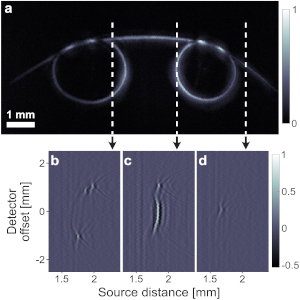
Fig. PTAM pressure measurements. (a) Transmission spectrum of π-BG at different static pressures. The legend notes the pressure in kPa. (b) Normalized power transmission, (c) peak transmission, (d) resonance width, and (e) resonance wavelength of π-BG at different pressure calculated from the spectrum plotted in panel (a). (f) Schematic configuration of simultaneous ultrasound signal detection. Ultrasound signal, generated by a transducer, impinges the detection array at an angle. The setup results in a slight delay difference of the ultrasound signal along the detector array. (g) and (h) Measured ultrasound signals of the setup in panel (f), for resonators presented in Figs. 1(e) and 1(f), respectively.

Yoav Hazan, Michael Nagli, Ahiad Levi, and Amir Rosenthal

© 2022 Optica Publishing Group Optics Letters Vol. 47, Issue 21, pp. 5660-5663 (2022) https://doi.org/10.1364/OL.467652https://doi.org/10.1038/s44172-022-00030-7

Burst-mode pulse interferometry for enabling low-noise multi-channel optical detection of ultrasound.

A schematic drawing of BM-PI.

© 2022 Optica Publishing Group under the terms of the Optica Open Access Publishing Agreement© 2019 Optics Express, OSA publishing.

Abstract
Ultrasound detection via optical resonators can achieve high levels of miniaturization and sensitivity as compared to piezoelectric detectors, but its scale-up from a single detector to an array is highly challenging. While the use of wideband sources may enable parallel interrogation of multiple resonators, it comes at the cost of reduction in the optical power, and ultimately in sensitivity, per channel. In this work we have developed a new interferometric approach to overcome this signal loss by using high-power bursts that are synchronized with the time window in which ultrasound detection is performed. Each burst is composed of a train of low-noise optical pulses which are sufficiently wideband to interrogate an array of resonators with non-overlapping spectra. We demonstrate our method, termed burst-mode pulse interferometry, for interrogating a single resonator in which the optical power was reduced to emulate the power loss per channel that occurs in parallel interrogation of 20 to 200 resonators. The use of bursts has led to up 25-fold improvement in sensitivity without affecting the shape of the acoustic signals, potentially enabling parallel low-noise interrogation of resonator arrays with a single source.

[Read more…]

The optical signal at the CM (blue) and BM (red)

Fig. The optical signal at the CM (blue) and BM (red) of the CRF output in the transition from the unlocked stated, in which the CRF blocks the optical pulses, to the locked state in which the CRF transmits the optical pulses and blocks only the ASE. Each of the spikes in the locked BM channel represents a burst with a width of 10 µs, shown in detail in Fig. 2(d), where the burst repletion rate was 8 kHz.

Oleg Volodarsky, Yoav Hazan, Michael Nagli, and Amir Rosenthal.
Optics Express, Volume 30, Issue 6, Pages 8959-8973
https://opg.optica.org/oe/abstract.cfm?URI=oe-30-6-8959


Simultaneous multi-channel ultrasound detection via phase modulated pulse interferometry.

The acoustic setup used to test the performance of PM-PI.

© 2019 Optics Express, OSA publishing.

Abstract
In optical detection of ultrasound, resonators with high Q-factors are often used to maximize sensitivity. However, in order to perform parallel interrogation, conventional interferometric techniques require an overlap between the spectra of all the resonators, which is difficult to achieve with high Q-factor resonators. In this paper, a new method is developed for parallel interrogation of optical resonators with non-overlapping spectra. The method is based on a phase-modulation scheme for pulse interferometry (PM-PI) and requires only a single photodetector and sampling channel per ultrasound detector. Using PM-PI, parallel ultrasound detection is demonstrated with four high Q-factor resonators.

[Read more…]

A schematic drawing of the PM-PI.

Fig. A schematic drawing of the PM-PI system used in this work to interrogate 4 resonators, implemented with π-phase shifted fiber Bragg gratings (π-FBGs). A wideband pulse laser with band-pass filters (BPFs) and an erbium-doped fiber amplifier (EDFA) create a source with a high spectral power density and sufficient bandwidth to cover the spectra of all the resonators. The modulation unit is an unbalanced Mach-Zehnder interferometer (MZI), composed of optical fiber couplers (FC) and a phase modulator (PM). The input phase signal to the PM, shown in the top-right plot, alternates between two values with a difference of 𝜋/2. For each phase value, the pulses interfere differently at the output of each resonator depending on the phase difference in the MZI for the specific resonance wavelength of that resonator. The bottom-right plot, shows a typical voltage signal measured for one of the resonators, which alternates between two states that correspond to the two phase values. As the bottom-right plot shows, in the current implementation, the duration of each phase value delivered to the PM corresponded to 5 laser pulses. We note that the limited bandwidth of our measurement did not allow full separation between the pulses in the bottom-right plot.

Yoav Hazan and Amir Rosenthal. 
Optics Express Vol. 27, Issue 20, pp. 28844-28854 (2019) https://doi.org/10.1364/OE.27.028844


Enhanced Sensitivity of Silicon-Photonics-Based Ultrasound Detection via BCB Coating.

Sio and Bcb over clading

© 2019 IEEE Photonics Journal

Impact Statement:
This paper gives a solution to one of the fundamental limitations of silicon-photonics based ultrasound detectors: the low photo-elastic response of silicon and silica. By using a BCB over-cladding, 5-fold increase in the acoustic sensitivity is achieved. We additionally provide a detailed analysis of the sensing mechanism, quantifying the different effects that contribute to the enhanced sensitivity.
Abstract:
Ultrasound detection via silicon waveguides relies on the ability of acoustic waves to modulate the effective refractive index of the guided modes. However, the low photo-elastic response of silicon and silica limits the sensitivity of conventional silicon-on-insulator sensors, in which the silicon core is surrounded by a silica cladding. In this paper, we demonstrate that the sensitivity of silicon waveguides to ultrasound may be significantly enhanced by replacing the silica over-cladding with bisbenzocyclobutene (BCB)-a transparent polymer with a high photo-elastic coefficient. In our experimental study, the response to ultrasound, in terms of the induced modulation in the effective refractive index, achieved for a BCB-coated silicon waveguide with TM polarization was comparable to values previously reported for polymer waveguides and an order of magnitude higher than the response achieved by an optical fiber. In addition, in our study, the susceptibility of the sensors to surface acoustic waves and reverberations was reduced for both TE and TM modes when the BCB over-cladding was used.

[Read more…]

setup schematic

Fig. – The measurement setup. For each of the polarizations, a Mach-Zehnder interferometer was constructed, where in each of the interferometer arms a chip with a different over-cladding material (BCB or silica) was connected. An ultrasound transducer was used to generate acoustic waves that impinged on only one of the chips, which were separated by more than 10 cm.

Resmi Ravi Kumar, Evgeny Hahamovich, Shai Tsesses, Yoav Hazan, Assaf Grinberg, Amir Rosenthal. IEEE Photonics Journal ( Volume: 11 , Issue: 3 , June 2019 )

Noise reduction in resonator-based ultrasound sensors by using a CW laser and phase detection.

© 2019 Optical Society of America

The detection of ultrasound via optical resonators is conventionally performed by tuning a continuous-wave (CW) laser to the linear slope of the resonance and monitoring the intensity modulation at the resonator output. While intensity monitoring offers the advantage of simplicity, its sensitivity is often limited by the frequency noise of the CW laser. In this work, we develop an alternative CW technique that can significantly reduce measurement noise by monitoring variations in the phase, rather than intensity, at the resonator output. In our current implementation, which is based on a balanced Mach–Zehnder interferometer for phase detection, we demonstrate a 24-fold increase in the signal-to-noise ratio of the detected ultrasound signal over the conventional, intensity-monitoring approach.[Read more…]

Signals detected using the IM and PM scheme

Fig. – Signals detected using the IM and PM interrogation methods with an optical power of 0.1 mW. In the PM measurement, the OPD was set to zero to maximize the SNR.

Lucas Riobó, Yoav Hazan, Francisco Veiras, María Garea, Patricio Sorichetti, and Amir Rosenthal.  Optics Letters Vol. 44, Issue 11, pp. 2677-2680 (2019) •https://doi.org/10.1364/OL.44.002677
© 2019 Optical Society of America

Ultrasound detection via low-noise pulse interferometry using a free-space Fabry-Pérot

source: © 2018 Optical Society of America

Coherence-restored pulse interferometry (CRPI) is a recently developed method for optical detection of ultrasound that achieves shot-noise-limited sensitivity and high dynamic range. In principle, the wideband source employed in CRPI may enable the interrogation of multiple detectors by using wavelength multiplexing. However, the noise-reduction scheme in CRPI has not been shown to be compatible with wideband operation. In this work, we introduce a new scheme for CRPI that relies on a free-space Fabry-Pérot filter for noise reduction and a pulse stretcher for reducing nonlinear effects. Using our scheme, we demonstrate that shot-noise-limited detection may be achieved for a spectral band of 80 nm and powers of up to 5 mW. [Read More…]

Fig. 1 A schematic of CRPI. EDFA is erbium-doped fiber amplifier; PZ is piezoelectric fiber stretcher; CRF is coherence-restoring filter; and π-FBG is π-phase-shifted fiber Bragg grating. The pulse train from the laser is filtered to a bandwidth of 0.4 nm, amplified, and further filtered by the CRF. Shifts of resonance of the π-FBG are measured by optical demodulator, implemented by a Mach-Zehnder interferometer locked to quadrature.

Oleg Volodarsky, Yoav Hazan, and Amir Rosenthal, “Ultrasound detection via low-noise pulse interferometry using a free-space Fabry-Pérot,” Opt. Express 26, 22405-22418 (2018)